Lung cancer
Non-small cell lung cancer TNM definitions (UICC update 1997)
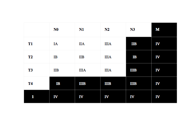
The following are the TNM definitions for non-small cell lung cancer adopted by the UICC and AJCC in 1997. These tumours are then grouped into Stages I ,II, III and IV (see diagram below).
Primary tumor (T)
TX: Primary tumor cannot be assessed, or tumor proven by the presence of malignant cells in sputum or bronchial washings but not visualised by imaging or bronchoscopy
T0: No evidence of primary tumour
TIS: Carcinoma in situ
T1: A tumour that is 3 cm or less in greatest dimension, surrounded by lung or visceral pleura, and without bronchoscopic evidence of invasion more proximal than the lobar bronchus (i.e., not in a main bronchus)*
T2: A tumour with any of the following features of size or extent:
-
•More than 3 cm in greatest dimension
-
•Involves a main bronchus, 2 cm or more distal to the carina
-
•Invades the visceral pleura
-
•Associated with atelectasis or obstructive pneumonitis that extends to the hilar region but does not involve the entire lung
T3: A tumour of any size that directly invades any of the following:
-
•chest wall (including superior sulcus tumours), diaphragm, mediastinal pleura (or phrenic nerve), parietal pericardium; or
-
•tumour in the main bronchus less than 2 cm distal to the carina but without involvement of the carina; or
-
•associated atelectasis or obstructive pneumonitis of the entire lung
T3a: atelectasis or obstructive pneumonitis of one entire lung without other criterion for T3.
T3b: Other criterion present for T3
T4: A tumour of any size that invades any of the following:
-
•mediastinum, heart, great vessels, trachea, oesophagus, recurrent laryngeal nerve, vertebral body, carina; or
-
•separate tumour nodules in the same lobe; or
-
•tumour with a malignant pleural effusion **
T4a: all T4 except T4b
T4b: invasion of the carina or presence of a malignant pleural effusion
*Note: The uncommon superficial tumour of any size with its invasive component limited to the bronchial wall, which may extend proximal to the main bronchus, is also classified as T1.
**Note: Most pleural effusions associated with lung cancer are due to tumour. However, there are a few patients in whom multiple cytopathologic examinations of pleural fluid are negative for tumour. In these cases, fluid is non-bloody and is not an exudate. When these elements and clinical judgement dictate that the effusion is not related to the tumour, the effusion should be excluded as a staging element and the patient should be staged as T1, T2, or T3.
Regional lymph nodes (N)
NX: Regional lymph nodes cannot be assessed
N0: No regional lymph node metastasis
N1: Metastasis to ipsilateral peribronchial and/or ipsilateral hilar lymph nodes, and intrapulmonary nodes including involvement by direct extension of the primary tumour
N2: Metastasis to ipsilateral mediastinal and/or subcarinal lymph node(s)
-
•N2a: Metastases in ipsilateral mediastinal nodes other than the paratracheal or para-oesophageal nodes
-
•N2b: Metastases in the paratracheal and para-oesophageal nodes
N3: Metastasis to contralateral mediastinal, contralateral hilar, ipsilateral or contralateral scalene, or supraclavicular lymph node(s)
-
•N3a: Metastases in the contralateral (hilar or mediastinal) nodes
-
•N3b: Metastases in the supra-clavicular fossae or scalene nodes
Distant metastasis (M)
MX: Distant metastasis cannot be assessed
M0: No distant metastasis
M1: Distant metastasis present
Note: M1 includes separate tumour nodule(s) in a different lobe (ipsilateral or contralateral).
Specify sites according to the following notations:
BRA = brain EYE = eye HEP = hepatic
LYM = lymph nodes MAR = bone marrow OSS = osseous
OTH = other OVR = ovary PER = peritoneal
PLE = pleura PUL = pulmonary SKI = skin