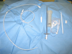
The Pleur-X catheter comes in a ready packed kit consisting of: Aspiration needle and syringe, guidewire, tunneller, cuffed catheter, introducing sheath, aspiration tubing, sterile cap and bandaging.

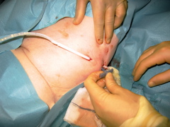
After injection of local anaesthetic a 1cm incision is made at the site of catheter insertion. Effusion is confirmed by aspiration.
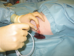
The Seldinger guidewire is inserted through the needle.
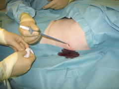
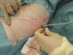
The introducer is passed over the wire.
The cuffed catheter is attached to the tunneller.
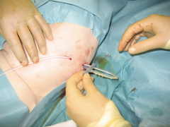
A second incision is made at the position where the catheter will egress the skin. It is best to try to place this relatively anteriorly so that aspiration and dressing can be performed easily. The catheter is then tunnelled to the initial incision alongside the introducing sheath.
The cuff is placed just under the skin.
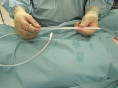
The catheter is then fed into the introducing sheath.
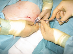
Holding the catheter in position with a mosquito forceps, the introducing sheath is split (breaking the tabs apart allows it to split easliy along pre-cut lines.
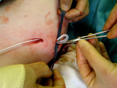
The introducing sheath is gradually removed.
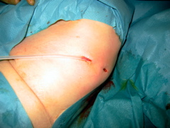
Any kinks are straightened to allow the catheter to lie smoothly under the skin. The incisions are closed with subcuticular sutures. Frequently the anterior incision needs no suture as long as it hasn't been maqde too large.
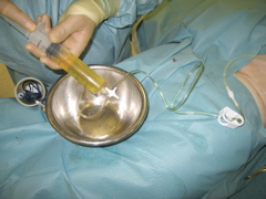
The effusion is drained to dryness before being capped and placed under dressings. The patient can be sent home the same day. Before discharge our ward nursing staff and MacMillan cancer nurses teach the district nurse how to apply the pre-vaccuumed cannister. Usually weekly aspiration is required. After 6-8 weeks the aspirate usually decreases and a form of pleurodesis is frequently achieved. The catheter can be removed under local anaesthetic.