From rudimentary beginnings ... to complete service.
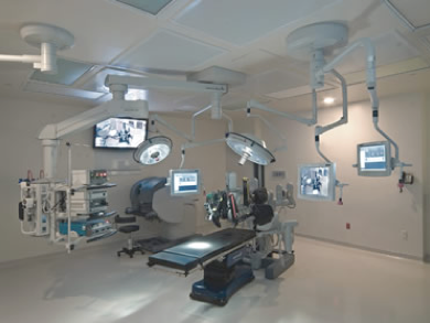
Some surgeons would have you believe that you need a fully equipped endoscopic surgical suite, equipped with operating lights, cameras, multiple TV screens all on overhead booms and a fully equipped surgical robot. However when we performed our first thoracoscopic lung biopsy we had no camera, no TV screens, no carbon dioxide insufflation and there was no assistant. The sales representative from the stapler company assisted while I looked down the optical telescope which was held by the scrub nurse and stapled off the affected part of the lung with the early surgical staplers. As you can see the only equipment used was a small Duval forceps, a straight 10mm optical telescope and a surgical stapler. While I thought that we would never be able to do much with this ‘keyhole’ approach, within weeks with this minimal equipment we were able to perform pleural biopsies, blebectomy, talc pleurodesis, lysis of adhesions and lung biopsies.
However the use of a video camera has opened up the ability to perform a full spectrum of thoracic procedures. As the assistants and scrub team can all see the action on the video camera, it is possible not only for the surgeon to operate but for the assistants to actively be involved with the operation. Most thoracoscopic procedures are in fact “two operator” procedures with “the assistant” playing a major part in the procedure.
VATS lobectomy represents a more advanced level of minimal access surgery and is probably one of the more intricate endoscopic procedures that can be performed. I did not have the confidence that a VATS approach could provide a proper oncological clearance till we had access to PET scanning. Where there is a small peripheral lesion and the PET shows no hilar or mediastinal uptake, I am now prepared to perform lobectomy.
As one becomes more experienced the whole range of thoracoscopic procedures becomes possible. We have performed the following procedures now thoracoscopically:
-
-
•VATS pneumonectomy
-
•VATS anatomical segmentectomy
-
•VATS lung volume reduction
-
•VATS decortication of empyema
-
•VATS excision of neurogenic tumour of the mediastinum
-
•VATS thymectomy
-
•VATS mobilisation of oesophagus for oesophagectomy
-
•VATS excision of pericardial and dermoid mediastinal cysts.
-
-
Having become adept at the above procedures it is clear that there a number of other procedures which can be performed using a thoracoscope:
-
-
1.VATS ablation of atrial fibrillation foci – the “mini-maze” type procedure
-
2.VATS harvest of IMA with off-pump graft to LAD
-
3.VATS first rib resection for thoracic outlet syndrome
-
4.VATS correction of scoliosis
-
-
While we have not yet performed these procedures, it is our aim to do so in the near future with the assistance of our cardiac and orthopaedic colleagues.