<--Previous Up Next-->
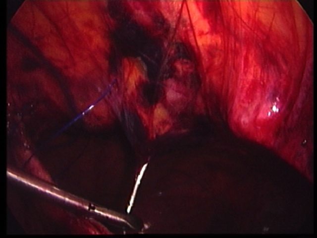
As no muscular diaphragmatic remnant exists anteriorly into which one can reliably place a suture or fix a patch, the plan was to suture the edge of the diaphragm to definitive anterior abdominal wall structures, burying the knots in small cutaneous incisions along the costal margin. Interrupted 2/0 Prolene sutures on a straightened needle were passed through the anterior abdominal wall. The terminal branches of the internal vessels can be seen in the right limit of the hernia. The xiphisternum is seen projecting into the middle of the defect to the right side of this photo. The left mammary vessels passed through the conralateral limit of the defect confirming that a Morgagni hernia is indeed a midline hernia.